 by
by By Dr. Amit Tandon – Dr. Kamlesh Tandon Hospital, IVF Center and Robotic Surgery Center, Agra
Introduction
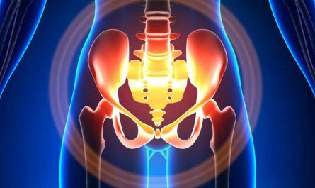
Endometrial cancer is the most common gynecological malignancy in developed countries. Accurate diagnosis at an early stage is crucial for effective treatment and improved survival. A follow‑up hysteroscopy with biopsy is a key diagnostic procedure that allows direct visualization of the uterine cavity and targeted tissue sampling for histopathological evaluation.
Why Hysteroscopy + Biopsy is Important
- Direct Visualization – Enables identification of lesions, polyps, or abnormal vascular patterns that may not be seen on imaging alone.
- Targeted Biopsy – Improves diagnostic accuracy by sampling suspicious areas under direct vision, reducing false‑negative rates.
- Minimally Invasive – Performed as an outpatient procedure with a small flexible scope, offering quick recovery.
Typical Indications for Follow‑Up Hysteroscopy
- Abnormal uterine bleeding (post‑menopausal, irregular, or heavy).
- Thickened endometrium (>4–5 mm on ultrasound) or suspicious endometrial lesions on imaging.
- Persistent endometrial hyperplasia on prior biopsy.
- Surveillance in high‑risk patients (e.g., Lynch syndrome, tamoxifen users).
Procedure Overview (Step‑by‑Step)
Pre‑Procedure
- Informed consent – Discuss risks (bleeding, infection, uterine perforation) and benefits.
- Pre‑operative evaluation – CBC, coagulation profile, ultrasound to assess endometrial thickness.
- Timing – Preferably after menstrual cycle (if applicable) or post‑menopause at any time.
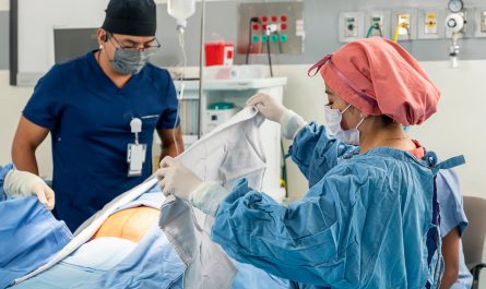
During Hysteroscopy
- Positioning – Dorsal lithotomy position; sterile draping.
- Cervical dilation – Gentle dilation if needed for scope insertion.
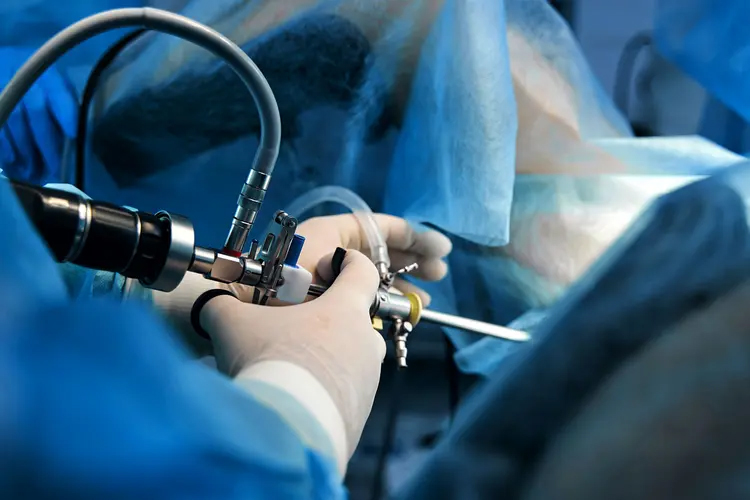
- Hysteroscope insertion – A thin, flexible or rigid hysteroscope with saline or CO₂ distension to visualize the uterine cavity.
- Systematic inspection – Evaluate endometrial lining, tubal ostia, and any abnormal areas.
- Targeted biopsy – Use a biopsy grasper or Pipelle device to obtain multiple samples from suspicious lesions or representative areas.
Post‑Procedure
- Observe for 15–30 min for any immediate complications (excessive bleeding, pain).
- Provide discharge instructions: avoid intercourse/tampons for 1–2 weeks, report heavy bleeding or fever.
- Histopathology report usually available within 5–7 days.
Diagnostic Accuracy
- Sensitivity of hysteroscopy‑guided biopsy for endometrial cancer approaches 95–99% when combined with histology.
- Specificity is high (~90%) for differentiating benign vs. malignant lesions.
- Reference: Novak’s Gynecology, 16th ed., p. 482 (details on diagnostic accuracy of hysteroscopy).
Role of Hysteroscopy in Endometrial Cancer Management
Stage Use of Hysteroscopy
Diagnosis Confirms malignancy, determines tumor location & extent.
Pre‑operative planning Assists in assessing myometrial invasion, cervical involvement.
Follow‑up Monitors response to treatment, detects recurrence.
Advantages of Performing at Dr. Kamlesh Tandon Hospital
- Expertise of Dr. Amit Tandon – Over 20+ years of experience in advanced hysteroscopic procedures and gynecologic oncology.
- State‑of‑the‑art hysteroscopy suite with high‑definition imaging and CO₂ insufflation for clear visualization.
- On‑site pathology lab – Rapid processing and reporting of biopsy specimens.
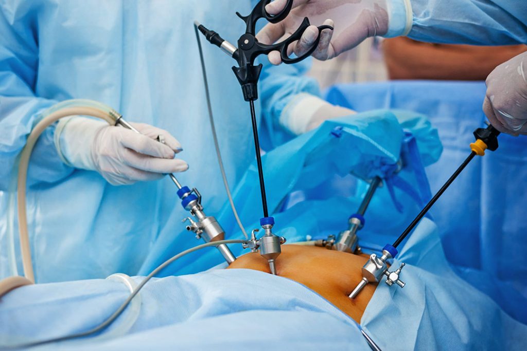
- Robotic surgical backup – If a more extensive surgery (e.g., hysterectomy) is required later, our robotic team is available.
Post‑Biopsy Management (If Endometrial Cancer is Diagnosed)
- Staging work‑up – MRI pelvis, CT chest/abdomen, CA‑125.
- Multidisciplinary discussion – Tumor board review for treatment planning (surgery, radiation, chemotherapy).
- Surgical options – Total laparoscopic or robotic hysterectomy with bilateral salpingo‑oophorectomy (often preferred at our center for faster recovery).
References (Books with Page Numbers)
- Novak’s Gynecology – 16th edition, p. 482 (Hysteroscopy diagnostic accuracy).
- Williams Gynecology – 4th edition, p. 1123 (Endometrial cancer work‑up).
- Berek & Novak’s Gynecology – 16th edition, p. 1345 (Hysteroscopic evaluation of abnormal uterine bleeding).
- Clinical Gynecologic Oncology – 9th edition, p. 210 (Role of hysteroscopy in endometrial cancer staging).
Hospital Details & Contact
Dr. Kamlesh Tandon Hospital
IVF Center and Robotic Surgery Center, Agra
We are dedicated to providing precise diagnosis and compassionate care for all our patients.